Molecular imaging

Molecular imaging is a field of medical imaging that focuses on imaging molecules of medical interest within living patients. This is in contrast to conventional methods for obtaining molecular information from preserved tissue samples, such as histology. Molecules of interest may be either ones produced naturally by the body, or synthetic molecules produced in a laboratory and injected into a patient by a doctor. The most common example of molecular imaging used clinically today is to inject a contrast agent (e.g., a microbubble, metal ion, or radioactive isotope) into a patient's bloodstream and to use an imaging modality (e.g., ultrasound, MRI, CT, PET) to track its movement in the body. Molecular imaging originated from the field of radiology from a need to better understand fundamental molecular processes inside organisms in a noninvasive manner.

The ultimate goal of molecular imaging is to be able to noninvasively monitor all of the biochemical processes occurring inside an organism in real time. Current research in molecular imaging involves cellular/molecular biology, chemistry, and medical physics, and is focused on: 1) developing imaging methods to detect previously undetectable types of molecules, 2) expanding the number and types of contrast agents available, and 3) developing functional contrast agents that provide information about the various activities that cells and tissues perform in both health and disease.

Overview

Molecular imaging emerged in the mid twentieth century as a discipline at the intersection of molecular biology and in vivo imaging. It enables the visualisation of the cellular function and the follow-up of the molecular process in living organisms without perturbing them. The multiple and numerous potentialities of this field are applicable to the diagnosis of diseases such as cancer, and neurological and cardiovascular diseases. This technique also contributes to improving the treatment of these disorders by optimizing the pre-clinical and clinical tests of new medication. They are also expected to have a major economic impact due to earlier and more precise diagnosis. Molecular and Functional Imaging has taken on a new direction since the description of the human genome. New paths in fundamental research, as well as in applied and industrial research, render the task of scientists more complex and increase the demands on them. Therefore, a tailor-made teaching program is in order.

Molecular imaging differs from traditional imaging in that probes known as biomarkers are used to help image particular targets or pathways. Biomarkers interact chemically with their surroundings and in turn alter the image according to molecular changes occurring within the area of interest. This process is markedly different from previous methods of imaging which primarily imaged differences in qualities such as density or water content. This ability to image fine molecular changes opens up an incredible number of exciting possibilities for medical application, including early detection and treatment of disease and basic pharmaceutical development. Furthermore, molecular imaging allows for quantitative tests, imparting a greater degree of objectivity to the study of these areas. One emerging technology is MALDI molecular imaging based on mass spectrometry.[citation needed]

Many areas of research are being conducted in the field of molecular imaging. Much research is currently centered on detecting what is known as a predisease state or molecular states that occur before typical symptoms of a disease are detected. Other important veins of research are the imaging of gene expression and the development of novel biomarkers. Organizations such as the SNMMI Center for Molecular Imaging Innovation and Translation (CMIIT) have formed to support research in this field. In Europe, other "networks of excellence" such as DiMI (Diagnostics in Molecular Imaging) or EMIL (European Molecular Imaging Laboratories) work on this new science, integrating activities and research in the field. In this way, a European Master Programme "EMMI" is being set up to train a new generation of professionals in molecular imaging.

Recently the term molecular imaging has been applied to a variety of microscopy and nanoscopy techniques including live-cell microscopy, Total Internal Reflection Fluorescence (TIRF)-microscopy, STimulated Emission Depletion (STED)-nanoscopy and Atomic Force Microscopy (AFM) as here images of molecules are the readout.

Imaging modalities

There are many different modalities that can be used for noninvasive molecular imaging. Each have their different strengths and weaknesses and some are more adept at imaging multiple targets than others.

Magnetic resonance imaging

Molecular MRI of a mouse brain presenting acute inflammation in the right hemisphere. Whereas unenhanced MRI failed to reveal any difference between right en left hemispheres, injection of a contrast-agent targeted to inflamed vessels allows to reveal inflammation specifically in the right hemisphere.

MRI has the advantages of having very high spatial resolution and is very adept at morphological imaging and functional imaging. MRI does have several disadvantages though. First, MRI has a sensitivity of around 10−3 mol/L to 10−5 mol/L which, compared to other types of imaging, can be very limiting. This problem stems from the fact that the difference between atoms in the high energy state and the low energy state is very small. For example, at 1.5 Tesla, a typical field strength for clinical MRI, the difference between high and low energy states is approximately 9 molecules per 2 million.[citation needed] Improvements to increase MR sensitivity include increasing magnetic field strength, and hyperpolarization via optical pumping, dynamic nuclear polarization or parahydrogen induced polarization. There are also a variety of signal amplification schemes based on chemical exchange that increase sensitivity.[1]

To achieve molecular imaging of disease biomarkers using MRI, targeted MRI contrast agents with high specificity and high relaxivity (sensitivity) are required. To date, many studies have been devoted to developing targeted-MRI contrast agents to achieve molecular imaging by MRI. Commonly, peptides, antibodies, or small ligands, and small protein domains, such as HER-2 affibodies, have been applied to achieve targeting. To enhance the sensitivity of the contrast agents, these targeting moieties are usually linked to high payload MRI contrast agents or MRI contrast agents with high relaxivities.[2] In particular, the recent development of micron-sized particles of iron oxide (MPIO) allowed to reach unprecedented levels of sensitivity to detect proteins expressed by arteries and veins.[3]

Optical imaging

Imaging of engineered E. coli Nissle 1917 in the mouse gut

There are a number of approaches used for optical imaging. The various methods depend upon fluorescence, bioluminescence, absorption or reflectance as the source of contrast.[4]

Optical imaging's most valuable attribute is that it and ultrasound do not have strong safety concerns like the other medical imaging modalities.[citation needed]

The downside of optical imaging is the lack of penetration depth, especially when working at visible wavelengths. Depth of penetration is related to the absorption and scattering of light, which is primarily a function of the wavelength of the excitation source. Light is absorbed by endogenous chromophores found in living tissue (e.g. hemoglobin, melanin, and lipids). In general, light absorption and scattering decreases with increasing wavelength. Below ~700 nm (e.g. visible wavelengths), these effects result in shallow penetration depths of only a few millimeters. Thus, in the visible region of the spectrum, only superficial assessment of tissue features is possible. Above 900 nm, water absorption can interfere with signal-to-background ratio. Because the absorption coefficient of tissue is considerably lower in the near infrared (NIR) region (700-900 nm), light can penetrate more deeply, to depths of several centimeters.[5]

Near Infrared imaging

Fluorescent probes and labels are an important tool for optical imaging. Some researchers have applied NIR imaging in rat model of acute myocardial infarction (AMI), using a peptide probe that can binds to apoptotic and necrotic cells.[6] A number of near-infrared (NIR) fluorophores have been employed for in vivo imaging, including Kodak X-SIGHT Dyes and Conjugates, Pz 247, DyLight 750 and 800 Fluors, Cy 5.5 and 7 Fluors, Alexa Fluor 680 and 750 Dyes, IRDye 680 and 800CW Fluors. Quantum dots, with their photostability and bright emissions, have generated a great deal of interest; however, their size precludes efficient clearance from the circulatory and renal systems while exhibiting long-term toxicity.[citation needed].

Several studies have demonstrated the use of infrared dye-labeled probes in optical imaging.

  1. In a comparison of gamma scintigraphy and NIR imaging, a cyclopentapeptide dual-labeled with 111
    In
    and an NIR fluorophore was used to image αvβ3-integrin positive melanoma xenografts.[7]
  2. Near-infrared labeled RGD targeting αvβ3-integrin has been used in numerous studies to target a variety of cancers.[8]
  3. An NIR fluorophore has been conjugated to epidermal growth factor (EGF) for imaging of tumor progression.[9]
  4. An NIR fluorophore was compared to Cy5.5, suggesting that longer-wavelength dyes may produce more effective targeting agents for optical imaging.[10]
  5. Pamidronate has been labeled with an NIR fluorophore and used as a bone imaging agent to detect osteoblastic activity in a living animal.[11]
  6. An NIR fluorophore-labeled GPI, a potent inhibitor of PSMA (prostate specific membrane antigen).[12]
  7. Use of human serum albumin labeled with an NIR fluorophore as a tracking agent for mapping of sentinel lymph nodes.[13]
  8. 2-Deoxy-D-glucose labeled with an NIR fluorophore.[14]

It is important to note that addition of an NIR probe to any vector can alter the vector's biocompatibility and biodistribution. Therefore, it can not be unequivocally assumed that the conjugated vector will behave similarly to the native form.

Single photon emission computed tomography

SPECT image (bone tracer) of a mouse MIP

The development of computed tomography in the 1970s allowed mapping of the distribution of the radioisotopes in the organ or tissue, and led to the technique now called single photon emission computed tomography (SPECT).

The imaging agent used in SPECT emits gamma rays, as opposed to the positron emitters (such as 18
F
) used in PET. There are a range of radiotracers (such as 99m
Tc
, 111
In
, 123
I
, 201
Tl
) that can be used, depending on the specific application.

Xenon (133
Xe
) gas is one such radiotracer. It has been shown to be valuable for diagnostic inhalation studies for the evaluation of pulmonary function; for imaging the lungs; and may also be used to assess rCBF. Detection of this gas occurs via a gamma camera—which is a scintillation detector consisting of a collimator, a NaI crystal, and a set of photomultiplier tubes.

By rotating the gamma camera around the patient, a three-dimensional image of the distribution of the radiotracer can be obtained by employing filtered back projection or other tomographic techniques. The radioisotopes used in SPECT have relatively long half lives (a few hours to a few days) making them easy to produce and relatively cheap. This represents the major advantage of SPECT as a molecular imaging technique, since it is significantly cheaper than either PET or fMRI. However it lacks good spatial (i.e., where exactly the particle is) or temporal (i.e., did the contrast agent signal happen at this millisecond, or that millisecond) resolution. Additionally, due to the radioactivity of the contrast agent, there are safety aspects concerning the administration of radioisotopes to the subject, especially for serial studies.

Positron emission tomography

Imaging joint inflammation in an arthritic mouse using positron emission tomography.
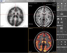
PET, MRI, and overlaid images of a human brain.

Positron emission tomography (PET) is a nuclear medicine imaging technique which produces a three-dimensional image or picture of functional processes in the body. The theory behind PET is simple enough. First a molecule is tagged with a positron emitting isotope. These positrons annihilate with nearby electrons, emitting two 511 keV photons, directed 180 degrees apart in opposite directions. These photons are then detected by the scanner, which can estimate the density of positron annihilations in a specific area. When enough interactions and annihilations have occurred, the density of the original molecule may be measured in that area. Typical isotopes include 11
C
, 13
N
, 15
O
, 18
F
, 64
Cu
, 62
Cu
, 124
I
, 76
Br
, 82
Rb
, 89
Zr
and 68
Ga
, with 18
F
being the most clinically utilized. One of the major disadvantages of PET is that most of the probes must be made with a cyclotron. Most of these probes also have a half life measured in hours, forcing the cyclotron to be on site. These factors can make PET prohibitively expensive. PET imaging does have many advantages though. First and foremost is its sensitivity: a typical PET scanner can detect between 10−11 mol/L to 10−12 mol/L concentrations.

See also

References

  1. ^ Gallagher, F.A. (2010). "An introduction to functional and molecular imaging with MRI". Clinical Radiology. 65 (7): 557–566. doi:10.1016/j.crad.2010.04.006. ISSN 0009-9260. PMID 20541655.
  2. ^ Shenghui, Xue; Jingjuan Qiao; Fan Pu; Mathew Cameron; Jenny J. Yang (17 Jan 2013). "Design of a novel class of protein-based magnetic resonance imaging contrast agents for the molecular imaging of cancer biomarkers". Wiley Interdiscip Rev Nanomed Nanobiotechnol. 5 (2): 163–79. doi:10.1002/wnan.1205. PMC 4011496. PMID 23335551.
  3. ^ Gauberti M, Montagne A, Quenault A, Vivien D (2014). "Molecular magnetic resonance imaging of brain-immune interactions". Front Cell Neurosci. 8: 389. doi:10.3389/fncel.2014.00389. PMC 4245913. PMID 25505871.
  4. ^ Weissleder R, Mahmood U (May 2001). "Molecular imaging". Radiology. 219 (2): 316–33. doi:10.1148/radiology.219.2.r01ma19316. PMID 11323453.
  5. ^ Kovar JL, Simpson MA, Schutz-Geschwender A, Olive DM (August 2007). "A systematic approach to the development of fluorescent contrast agents for optical imaging of mouse cancer models". Anal. Biochem. 367 (1): 1–12. doi:10.1016/j.ab.2007.04.011. PMID 17521598. as PDF Archived February 11, 2009, at the Wayback Machine
  6. ^ Acharya, B; Wang, K; Kim, IS; Kang, W; Moon, C; Lee, BH (2013). "In vivo imaging of myocardial cell death using a peptide probe and assessment of long-term heart function". Journal of Controlled Release. 172 (1): 367–73. doi:10.1016/j.jconrel.2013.08.294. PMID 24021357.
  7. ^ Houston JP, Ke S, Wang W, Li C, Sevick-Muraca EM (2005). "Quality analysis of in vivo near-infrared fluorescence and conventional gamma images acquired using a dual-labeled tumor-targeting probe". J Biomed Opt. 10 (5): 054010. Bibcode:2005JBO....10e4010H. doi:10.1117/1.2114748. PMID 16292970.
  8. ^ Chen K, Xie J, Chen X (2009). "RGD-human serum albumin conjugates as efficient tumor targeting probes". Mol Imaging. 8 (2): 65–73. doi:10.2310/7290.2009.00011. PMC 6366843. PMID 19397852. Archived from the original on 2014-03-26.
  9. ^ Kovar JL, Johnson MA, Volcheck WM, Chen J, Simpson MA (October 2006). "Hyaluronidase expression induces prostate tumor metastasis in an orthotopic mouse model". Am. J. Pathol. 169 (4): 1415–26. doi:10.2353/ajpath.2006.060324. PMC 1698854. PMID 17003496.
  10. ^ Adams KE, Ke S, Kwon S, et al. (2007). "Comparison of visible and near-infrared wavelength-excitable fluorescent dyes for molecular imaging of cancer". J Biomed Opt. 12 (2): 024017. Bibcode:2007JBO....12b4017A. doi:10.1117/1.2717137. PMID 17477732. S2CID 39806507.
  11. ^ Zaheer A, Lenkinski RE, Mahmood A, Jones AG, Cantley LC, Frangioni JV (December 2001). "In vivo near-infrared fluorescence imaging of osteoblastic activity". Nat. Biotechnol. 19 (12): 1148–54. doi:10.1038/nbt1201-1148. PMID 11731784. S2CID 485155.
  12. ^ Humblet V, Lapidus R, Williams LR, et al. (2005). "High-affinity near-infrared fluorescent small-molecule contrast agents for in vivo imaging of prostate-specific membrane antigen". Mol Imaging. 4 (4): 448–62. doi:10.2310/7290.2005.05163. PMID 16285907.
  13. ^ Ohnishi S, Lomnes SJ, Laurence RG, Gogbashian A, Mariani G, Frangioni JV (2005). "Organic alternatives to quantum dots for intraoperative near-infrared fluorescent sentinel lymph node mapping". Mol Imaging. 4 (3): 172–81. doi:10.1162/15353500200505127. PMID 16194449.
  14. ^ Kovar JL, Volcheck W, Sevick-Muraca E, Simpson MA, Olive DM (January 2009). "Characterization and performance of a near-infrared 2-deoxyglucose optical imaging agent for mouse cancer models". Anal. Biochem. 384 (2): 254–62. doi:10.1016/j.ab.2008.09.050. PMC 2720560. PMID 18938129. as PDF Archived July 13, 2011, at the Wayback Machine

Read other articles:

Asian pay television channel Television channel AXNCountrySingaporeBroadcast areaSoutheast Asia (including Singapore)NetworkKC Global Media AsiaHeadquartersNumber 10, Changi Business Park Central 2 #03-01, Hansapoint @ Changi Business Park, Changi, SingaporeProgrammingLanguage(s)EnglishMalayIndonesianMandarin ChineseTamilPicture format1080i HDTVOwnershipOwnerSony Pictures Entertainment (2004-2020) KC Global Media Asia (2020-present)Sister channelsAnimaxONEGEMHistoryLaunched21 September 1...


Bupati KotabaruPetahanaH. Sayed Jafar Al-Idrus, S.H.sejak 26 April 2021Masa jabatan5 tahunDibentuk1950Pejabat pertamaM. YamaniSitus webkotabarukab.go.id Berikut ini adalah daftar Bupati Kotabaru yang menjabat sejak pembentukannya pada tahun 1950. No Bupati Mulai Jabatan Akhir Jabatan Prd. Ket. Wakil Bupati 1 M. Yamani 1950 1951 1   – 2 Abdul Rasjid 1951 1955 2   3 Ibrahim Sedar 1955 1958 3   4 H.Abdul Muluk 1958 1959 4   5 H. A.Hudari 1960 1963 5   6 Basrindu 1...


English-Indian politician This article includes a list of general references, but it lacks sufficient corresponding inline citations. Please help to improve this article by introducing more precise citations. (January 2013) (Learn how and when to remove this template message) Nellie SenguptaNellie and Jatindra Mohan Sengupta on a 1985 stamp of India52nd President of the Indian National CongressIn office1933–1934Preceded byMadan Mohan MalaviyaSucceeded byRajendra Prasad Personal detailsBornE...
American politician (1832–1890) Orlow W. ChapmanChapman in 1900 publication5th United States Solicitor GeneralIn office1889–1890Appointed byBenjamin HarrisonPreceded byGeorge A. JenksSucceeded byWilliam Howard TaftMember of the New York Senatefrom the 24th districtIn office1868–1871Preceded byEzra CornellSucceeded byThomas I. Chatfield Personal detailsBorn(1832-01-07)January 7, 1832Ellington, Connecticut, U.S.DiedJanuary 19, 1890(1890-01-19) (aged 58)Washington, D.C., U.S.Resti...


United Arab Emirates Handball FederationUAEHFSportHandballOther sportsBeach handballOfficial websitewww.uaehandball.netAFFILIATIONSInternational federationIHFIHF member since1975Continental associationAHFNational Olympic CommitteeUnited Arab Emirates National Olympic Committee The United Arab Emirates Handball Federation (UAEHF) is an administrative body for handball and beach handball in United Arab Emirates. It is a member of the Asian Handball Federation (AHF) and the International Handbal...


Urse d'AbetotSheriff of WorcestershireIn officec. 1069–1108Preceded byCyneweard of Laughern[1]Succeeded byRoger d'AbetotRoyal constableIn officeafter 1087 – 1108 Personal detailsBornc. 1040Normandy, FranceDiedSummer of 1108SpouseAliceChildrenRoger d'Abetot, daughter (perhaps named Emmeline) Urse d'Abetot[a] (c. 1040 - 1108) was a Norman who followed King William I to England, and became Sheriff of Worcestershire and a royal official under him and...

American college football rivalry Kansas State–Nebraska football rivalry Kansas State Wildcats Nebraska Cornhuskers First meetingOctober 14, 1911Nebraska, 59–0Latest meetingOctober 7, 2010Nebraska, 48–13StatisticsMeetings total95All-time seriesNebraska leads, 78–15–2[1]Largest victoryNebraska, 59–0 (1911)Longest win streakNebraska, 29 (1969–1997) [Interactive fullscreen map + nearby articles] Locations of Kansas State and Nebraska The Kansas State–Nebraska football riv...


此條目需要补充更多来源。 (2020年9月17日)请协助補充多方面可靠来源以改善这篇条目,无法查证的内容可能會因為异议提出而被移除。致使用者:请搜索一下条目的标题(来源搜索:费利克斯·迪亚斯 — 网页、新闻、书籍、学术、图像),以检查网络上是否存在该主题的更多可靠来源(判定指引)。 费利克斯·迪亚斯Felix Diaz出生(1868-02-17)1868年2月17日墨西哥瓦哈卡州逝世...


  此條目介紹的是2008年的颱風辛樂克。关于其他同名的熱帶氣旋,请见「颱風辛樂克」。 颱風森拉克Typhoon Sinlaku(英文)颱風森拉克路徑圖颱風森拉克的路徑圖十分鐘平均風速颱風(JMA)185 km/h(100 kt)強烈颱風(CWB)185 km/h(51 m/s)超強颱風 (KMA)175 km/h(48 m/s)強颱風 (HKO)175 km/h颱風 (PAGASA)175 km/h二分鐘平均風速超强...

2006 American filmZzyzxPromotional posterDirected byRichard HalpernWritten byArt D'AlessandroProduced byRichard HalpernStarringKenny JohnsonRobyn CohenRyan FoxKayo ZepedaMusic byKays Al-AtrakchiProductioncompanyYarble.comRelease date February 4, 2006 (2006-02-04) Running time80 minutesCountryUnited StatesLanguageEnglishBudget$1,000,000 (estimated) Zzyzx is a 2006 thriller film produced and directed by Richard Halpern and starring Kenny Johnson, Robyn Cohen, Ryan Fox, and Kayo Z...


Taiwanese politician For other people named David Chou, see David Chou (disambiguation). David ChouChou Po-lunMLY周伯倫Member of the Legislative YuanIn office1 February 1993 – 30 January 2003ConstituencyTaipei County 1In office1 February 1993 – 31 January 1999ConstituencyTaipei CountyMember of the Taipei City CouncilIn office25 December 1986 – 31 January 1993 Personal detailsBorn (1954-11-13) 13 November 1954 (age 69)Taipei, TaiwanPolitical partyDemocr...


Branch of Rodnovery Part of a series onSlavic Native Faith Theory Theology and cosmology Rod Belobog-Chernobog Prav-Yav-Nav Triglav Svetovid Clusters of deities Identity and political philosophy Slavic Native Faith and Christianity Denominations Core denominations Authentism Bazhovism Ivanovism Kandybaism Levashovism Peterburgian Vedism Ringing Cedars' Anastasianism Slavic-Hill Rodnovery Sylenkoism Vseyasvetnaya Gramota Ynglism Not strictly related Assianism Blagovery Dead Water Institutions ...

Daddy's Home 2Poster resmiSutradara Sean Anders Produser Will Ferrell Adam McKay Chris Henchy John Morris Ditulis oleh Sean Anders John Morris Skenario Sean Anders John Morris BerdasarkanCharactersoleh Brian BurnsPemeran Will Ferrell Mark Wahlberg Linda Cardellini John Cena John Lithgow Mel Gibson Penata musikMichael AndrewsSinematograferJulio MacatPenyuntingBrad WilhitePerusahaanproduksiGary Sanchez ProductionsDistributorParamount PicturesTanggal rilis 10 November 2017 (2017-11-10...


Bagian dari seri tentangGereja Ortodoks TimurMosaik Kristos Pantokrator, Hagia Sofia Ikhtisar Struktur Teologi (Sejarah teologi) Liturgi Sejarah Gereja Misteri Suci Pandangan tentang keselamatan Pandangan tentang Maria Pandangan tentang ikon Latar belakang Penyaliban / Kebangkitan / KenaikanYesus Agama Kristen Gereja Kristen Suksesi apostolik Empat Ciri Gereja Ortodoksi Organisasi Otokefali Kebatrikan Batrik Ekumenis Tatanan keuskupan Klerus Uskup Imam Diakon Monastisisme Tingkatan ...


bip internal ribosome entry site (IRES)Predicted secondary structure and sequence conservation of IRES_BipIdentifiersSymbolIRES_BipAlt. SymbolsBip_IRESRfamRF00223Other dataRNA typeCis-reg; IRESDomain(s)EukaryotaGOGO:0043022SOSO:0000243PDB structuresPDBe The BiP internal ribosome entry site (IRES) is an RNA element present in the 5' UTR of the mRNA of BiP protein and allows cap-independent translation. BiP protein expression has been found to be significantly enhanced by the heat shock respons...

American judge (born 1974) Sarah GeraghtyJudge of the United States District Court for the Northern District of GeorgiaIncumbentAssumed office April 8, 2022Appointed byJoe BidenPreceded byAmy Totenberg Personal detailsBorn1974 (age 48–49)Chicago, Illinois, U.S.EducationNorthwestern University (BA)University of Michigan (MSW, JD) Sarah Elisabeth Geraghty (born 1974)[1] is an American lawyer from Georgia who serves as a United States district judge of the United States Di...


1983 passenger plane crash in Cuenca, Ecuador This article needs additional citations for verification. Please help improve this article by adding citations to reliable sources. Unsourced material may be challenged and removed.Find sources: 1983 TAME Boeing 737 crash – news · newspapers · books · scholar · JSTOR (May 2012) (Learn how and when to remove this template message) 1983 TAME Boeing 737 crashThe aircraft involved in the accident, photographed ...


This article needs additional citations for verification. Please help improve this article by adding citations to reliable sources. Unsourced material may be challenged and removed.Find sources: Kabul Library – news · newspapers · books · scholar · JSTOR (September 2014) (Learn how and when to remove this template message) Kabul Library is one of Afghanistan's oldest and largest libraries, located in the capital Kabul. It includes books in many languag...

British online gambling operator This article needs to be updated. Please help update this article to reflect recent events or newly available information. (May 2019) SportingbetTypeSubsidiaryIndustryGamblingFounded1997HeadquartersLondon, United KingdomKey peopleCEO Kenneth AlexanderProductsSports bettingFinancial bettingPoker (Paradise Poker)Casino (Paradise Casino)GamesBackgammonRevenue£1,577.2 million (2009)[1]Operating income£21.9 million (2009)[1]Net income£12.4 millio...


The Honourable DameCatherine TizardONZ, GCMG, GCVO, DBE, QSO, DStJGubernur Jenderal Selandia Baru ke-16Masa jabatan13 Desember 1990 – 21 Maret 1996Penguasa monarkiElizabeth IIPerdana MenteriJim BolgerPendahuluSir Paul ReevesPenggantiSir Michael Hardie BoysWali Kota Auckland ke-35Masa jabatan1983–1990WakilJohn Strevens (1983–86)Harold Goodman (1986–88)Phil Warren (1988–90)PendahuluColin KayPenggantiLes Mills Informasi pribadiLahirC...


Strategi Solo vs Squad di Free Fire: Cara Menang Mudah!